[REVEAL] Neuronal microscopy for cell behavioural examination and manipulation
![[REVEAL] Neuronal microscopy for cell behavioural examination and manipulation [REVEAL] Neuronal microscopy for cell behavioural examination and manipulation](/var/www_mchtr/storage/images/badania-i-nauka/projekty/projekty-aktualne/reveal-mikroskopia-neuronalna-do-badania-zachowan-komorek-i-manipulacji-nimi/13827-2-pol-PL/REVEAL-Neuronal-microscopy-for-cell-behavioural-examination-and-manipulation_medium.png)
head of the project: Wojciech Krauze, PhD
project duration: 01.01.2021-30.06.2025
grant assigned for the project: 5 984 600 EUR
funds assigned for the tasks at PW: 496 875 EUR
website: http://reveal-h2020.eu/
Dysfunctions in cellular homeostasis lay the foundations for numerous diseases and may take the form of incorrect structures, functions and behaviours in mammals cells. The heterogeneity phenotypes of cells initiating tumors is a characteristic feature of liver cancer, influencing its aggressiveness and resistance to treatment, making it the second cause of sickness worldwide. These differences are often too subtle to be detected or too rare, which stresses the urgent need for new biophotonics devices that can reliably discover these occurrences. To gain proofs of the processes that result in tissue heterogeniety causing liver cancer, as a part of the REVEAL project we propose to devise a “neutron microscope” - an instrument, in which the hardware and analytical software are flawlessly integrated and they utilise the neuron calculating networks to define the cells trajectory, i.e. the evolution of cells phenotypes. The software is to a large extent based on utilising neurons networks for creating images, analysing cells, predicting the fate of cells and make the decision for the following purposes:
- imaging and analysing the behaviour of thousands of cells at the same time in near real-time conditions;
- identifying the changes in cells conditions, indicating the disease origins;
- indicating localisation of an interesting cell, whereas the hardware will enable us to get a sample of selected cells for bioanalysis.
While imaging live cells provides information on the cell’s past, the molecular analysis will reflect its current biological state. The broad scope of our project is revolutionary - we imagine a future in which live cells microscopy and biomolecular analysis will constitute a continuum to generate a complex biologica timestamp for each cell we are interested in. Such a neuron microscope will be invaluable in identifying the mechanisms of disease onset.
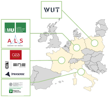
The project is executed as a part of a European consortium comprising of the following entities:
- Commissariat à l’énergie atomique et aux énergies alternatives (CEA), Francja
- IPRASENSE, Francja
- Ecole Normale Supérieure de Lyon (ENS Lyon), Francja
- Ludwig Maximilian University of Munich (LMU), Niemcy
- Fondazione IRCCS Ca’ Granda Ospedale Maggiore Policlinico Milano (PCLM), Włochy
- Automated Lab Solutions GmbH (ALS), Niemcy
- Politechnika Warszawska (WUT), Polska


